|
Surgeries involving Image Guidance Systems
(Orthopaedic Surgery, Neurosurgery, Spinal Surgery and ENT Surgery)
Cardiovascular Surgery
 
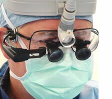
When performing surgery within the areas of the cranial cavity and spinal canal, it is of vital importance that neurosurgeons know the exact location of their surgical instruments. Using the MD-6 Critical Data Viewer, neurosurgeons view image-guidance data in real time to pinpoint the trajectory and tip location of their instruments as they use them ? without having to turn away to view the primary monitor.
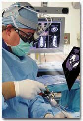 Due to the size of the cranial cavity and its lack of internal markers for localization, neurosurgeons rely on image-guidance systems for localizing and resecting tumors. By frequently referencing a MRI or CT scan with the MD-6, neurosurgeons determine exact instrument location and can thereby remove tumors in the cranial cavity more quickly and more accurately ? minimizing damage to surrounding critical brain structures. Due to the size of the cranial cavity and its lack of internal markers for localization, neurosurgeons rely on image-guidance systems for localizing and resecting tumors. By frequently referencing a MRI or CT scan with the MD-6, neurosurgeons determine exact instrument location and can thereby remove tumors in the cranial cavity more quickly and more accurately ? minimizing damage to surrounding critical brain structures.
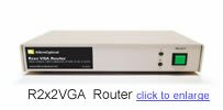 During spinal surgery, the MD-6 allows neurosurgeons to frequently view image guidance data in order to check the entry point for the pedicle screw as well as its trajectory during insertion. Reference to the viewer?s clear images within the surgeon?s field of view requires only eye movements which preserves hand-eye coordination. When the surgeon finds it necessary to view a digitally scanned x-ray, he can toggle between image-guidance scans and x-rays by using the R2x2 VGA Router. This eliminates the need for the surgeon to turn away to view the flouroscopy primary monitor. During spinal surgery, the MD-6 allows neurosurgeons to frequently view image guidance data in order to check the entry point for the pedicle screw as well as its trajectory during insertion. Reference to the viewer?s clear images within the surgeon?s field of view requires only eye movements which preserves hand-eye coordination. When the surgeon finds it necessary to view a digitally scanned x-ray, he can toggle between image-guidance scans and x-rays by using the R2x2 VGA Router. This eliminates the need for the surgeon to turn away to view the flouroscopy primary monitor.
| ?Image-guidance systems provide a major benefit to neurosurgeons. The MD-6 facilitates the accessing of this information which makes the system even more powerful by improving accuracy, operating speed, safety and ergonomics.? |
| ? Dr. James Macon, Surgical Neurology Associates |
To purchase products or to learn more about our medical partners, please contact us.

The MD-6 Critical Data Viewer allows cardiovascular surgeons to continually monitor a patient?s EKG traces, systemic blood pressure, pulmonary artery pressure, central venous pressure, core body temperature and SPO2. The MD-6 displays vitals right before the surgeons? eyes ? without obstructing their natural field of vision. Viewing vitals in real time without having to look away at a monitor allows surgeons to retain their concentration on the operative field.
 The user?s perception of the critical information displayed by the MD-6 is that of a floating image, similar to viewing a 13 inch monitor a few feet away. Small and lightweight in design, the viewer easily attaches to surgical and prescription eyewear. Using the R2x2 VGA Router, surgeons can toggle between 2 image sources to view 2 different sets of information, such as vital signs and x-ray images. The user?s perception of the critical information displayed by the MD-6 is that of a floating image, similar to viewing a 13 inch monitor a few feet away. Small and lightweight in design, the viewer easily attaches to surgical and prescription eyewear. Using the R2x2 VGA Router, surgeons can toggle between 2 image sources to view 2 different sets of information, such as vital signs and x-ray images.
�
�
�
|
?I believe MicroOptical?s viewers provide the next step in technology for the operative field and will revolutionize the way we do complex surgery.?
|
? Dr. Thomas Langdon, Cardiovascular Surgeon,
Omaha Cardiovascular and Thoracic Surgery |
To purchase products or to learn more about our medical partners, please contact us.

|